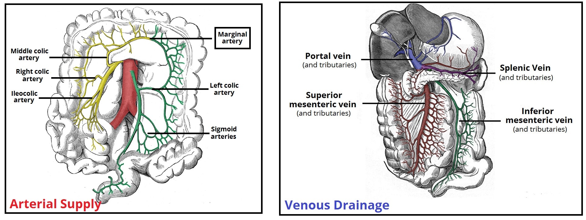
38 colon diagram with labels
Blog - Create a sequence diagram Labeling messages and roles in sequence diagrams Role names for instances of objects/systems and actors are indicated with a colon. E.g. : Customer Conditions (guards) on messages are indicated with square brackets either on the message, or in the frame shape. E.g. [value > $1000] Message numbers can be used to clearly show the sequence of events. Histology | Colon - The Common Vein This diagram illustrates the 4 basic layers of the colon. The inner pink layer is the mucosa, the yellow layer beneath the mucosa is called the submucosa, while the red layer is the muscular layer (muscularis) and the 4 th layer is called the serosa or adventitia. Courtesy Ashley Davidoff MD 32338 Ultrasound of normal large bowel
Colon Picture Labeling Flashcards | Quizlet Start studying Colon Picture Labeling. Learn vocabulary, terms, and more with flashcards, games, and other study tools. Search. Create. Log in Sign up. Upgrade to remove ads. ... ch.5 pathway of food diagram 14 Terms. olivia_ba. Digestive System Trace 28 Terms. lscott3. RBCs from mitral valve to tricuspid valve->traveling thru big toe 23 Terms ...

Colon diagram with labels
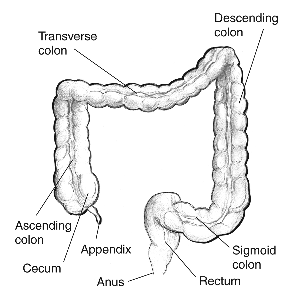
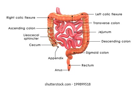
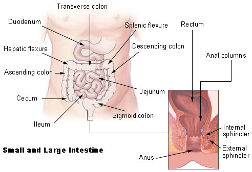
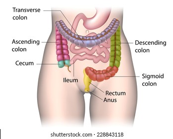
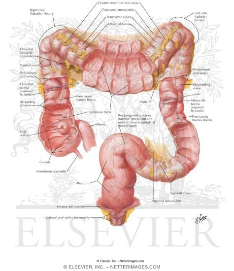
Illustration Picture of Anatomy - Colon - eMedicineHealth The colon is the largest part of the large intestine, extending from the cecum to the rectum. It is 5 feet long and its function is to reabsorb water from digested food and concentrate solid waste material, known as stool. The colon is made of several sections. The ascending colon travels up the right side of the abdomen. Colon Anatomy - Medical Art Library The colon is divided into four parts: the ascending, transverse, descendingand sigmoid. The ascending and transverse colon meet at the right hepatic flexure(near the liver). The transverse and descending colon meet at the left splenic flexure(near the spleen). The large intestine has three bands of longitudinal muscle fibers called taeniae coli. Abdomen and digestive system anatomy: diagrams labeled - IMAIOS Colon: three diagrams of the face of the colon and rectum, with or without the mesentery of the colon and taenia coli as well as a detailed illustration of the right colonic flexure. Large intestine
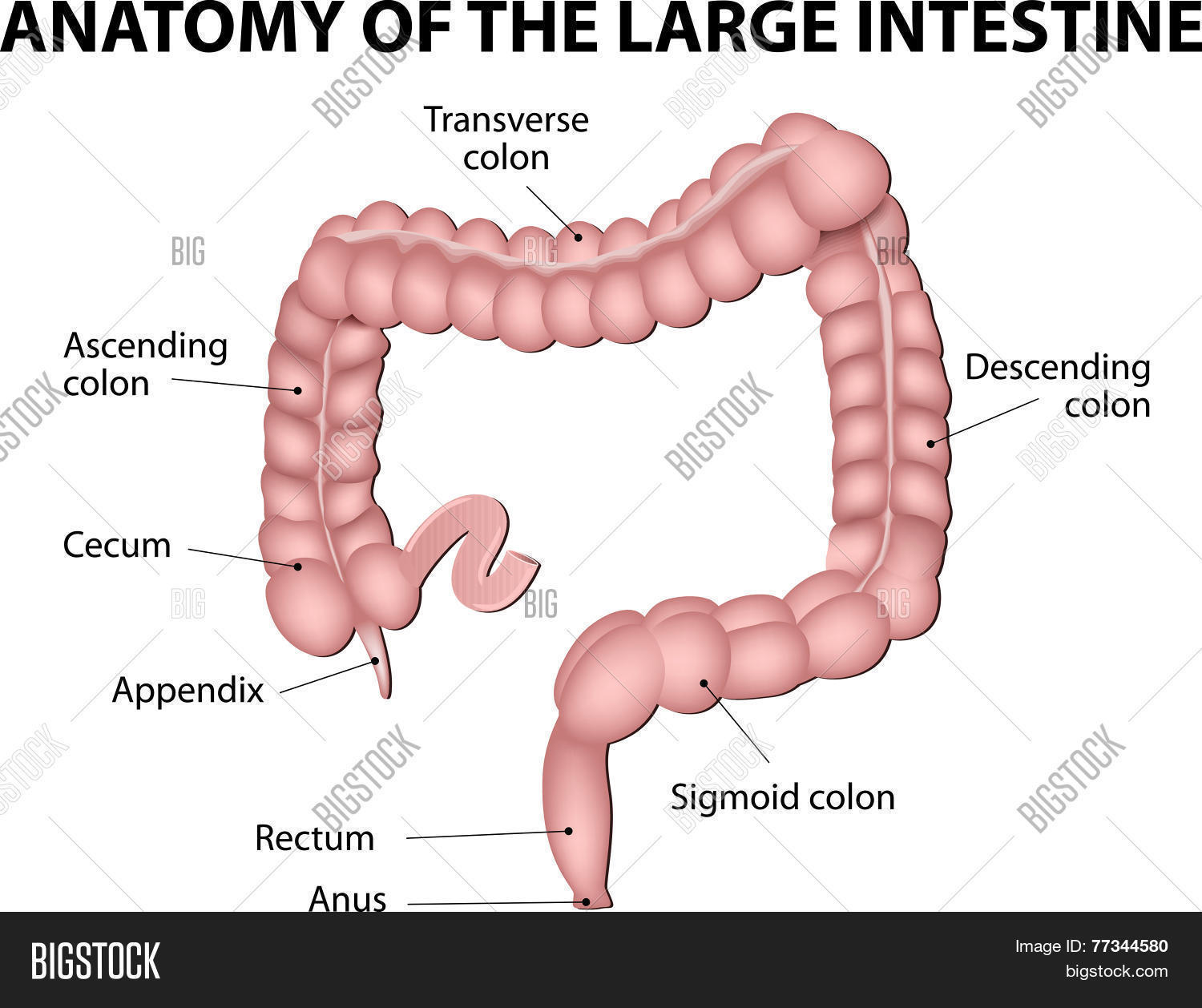
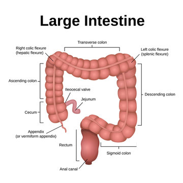
Colon diagram with labels. Understanding the Human Stomach Anatomy With Labeled Diagrams Given below is a labeled diagram of the stomach to help you understand stomach anatomy. The stomach is divided into four parts. These include: Cardia Fundus Body Pylorus Cardia refers to the section of the stomach that is located around the cardiac orifice. The lower esophageal sphincter lies at the junction where the esophagus meets the stomach. Table of Contents: - BYJUS Well-labelled Diagram of Large Intestine Large Intestine - Description The large intestine is a 1.5 m long organ that extends from the ileocaecal junction to the anus. It constitutes the cecum with an appendix, ascending colon, transverse colon, descending colon, pelvic or sigmoid colon, rectum and the anal canal. The Colon - Ascending - Transverse - Descending - Sigmoid - TeachMeAnatomy The colon (large intestine) is the distal part of the gastrointestinal tract, extending from the cecum to the anal canal. It receives digested food from the small intestine, from which it absorbs water and electrolytes to form faeces. Anatomically, the colon can be divided into four parts - ascending, transverse, descending and sigmoid. These sections form an arch, which encircles the small ... Label Digestive System Diagram Printout - EnchantedLearning.com Click here.) Read the definitions below, then label the digestive system anatomy diagram. anus - the opening at the end of the digestive system from which feces (waste) exits the body. appendix - a small sac located on the cecum. ascending colon - the part of the large intestine that run upwards; it is located after the cecum.
Diagram Of The Respiratory System With Labels Pictures, Images ... - iStock The colon and rectum are parts of the large intestine. 3D illustration diagram of the respiratory system with labels stock pictures, royalty-free photos & images Illustration of colon and rectum on transparent human model Colonoscopy Measurements (cm) from Anal Verge | SEER Training Types of Surgery: Colon; Types of Surgery: Rectum; Radiation Therapy; Commonly Used Drugs; For hands-on exercises, please go to SEER*Educate. Resources. Archived Modules. Updates. Acknowledgements. Colonoscopy Measurements (cm) from Anal Verge. Return to Anatomy of Colon and Rectum. Follow SEER. Contact Information. Human Intestines | Interactive Anatomy Guide - Innerbody Our large intestine consists of 4 major regions: the cecum, colon, rectum, and anal canal. The cecum is a pouch-like dead-end passage that branches inferiorly from the end of the ileum. Fecal matter entering the large intestine from the ileum passes into the cecum before being pushed superiorly into the ascending colon. The appendix is attached ... labeled engine diagram Honda Engine Gcv160 Parts Diagram - Best Diagram Collection . gcv160 gc160 hrr216vka mowers diagrams lawnmower mikrora navejas. 4 Toyota Tundra V4 Engine Diagram . camshaft schematic buharman. Andrews blog: four stroke engine. 4 toyota tundra v4 engine diagram. Colon diagram histology slides histological
How does the bowel work? Bowel information with diagrams Diagram of the position and sections of the small bowel The colon The colon is divided into 4 sections. Ascending colon The first part of the colon is joined to the small bowel and goes up the right side of the abdomen (tummy). Transverse colon The second section goes across the abdomen from your right to your left side. Descending colon Colon Anatomy (with Small Intestine Label) - National Cancer Institute 720x602. View. Download. Title: Colon Anatomy (with Small Intestine Label) Description: Drawing shows the cecum, ascending colon, transverse colon, descending colon, sigmoid colon, rectum, and anal canal. Also shown is the small intestine. The cecum connects the small intestine to the colon. Colon Diagram Illustrations & Vectors - Dreamstime Download 3,355 Colon Diagram Stock Illustrations, Vectors & Clipart for FREE or amazingly low rates! New users enjoy 60% OFF. 192,125,119 stock photos online. ... Labeled Diagram. Human colon. Colon cancer. 3d rendered illustration of a transparent female anatomy with tumor in colon. Female colon. 3d rendered illustration of a transparent ... Labeled Diagram of the Human Kidney - Bodytomy The renal medulla comprises a set of 8-18 conical structures called renal pyramids that are surrounded by the cortex. Portions of the cortex between two adjacent pyramids are termed as renal columns. Spread in these pyramids and the cortex, are the functional units callednephrons. The actual filtration of blood occurs in the nephrons.
Digestive system - AccessScience from McGraw-Hill Education
PDF Digestive System Diagram - direzionedidattica-vignola.edu.it Label these: #1 (Urine/pee pee) #2 (Solid Waste/poop) Hydrochloric Mechanical Digestion Chemical Digestion Saliva Acid Pepsin Bile Lipase Stomach Small Intestine Enzymes from Liver and Pancreas Large Intestine (Transverse Colon) Descending ColonCirculatory System Kidneys #1 #2 Water and Vitamins Nutrients The Digestive System

Labeled Large Intestine | CRS
Digestive organs: Diagram, stomach, intestines, and more The large intestine includes the cecum, transverse colon, ascending colon, descending colon, and sigmoid colon. A small, finger-like projection from the large intestine, the appendix, can become...

BIOL-2311L Large Intestine Anterior View Labeling Diagram ...
Intestines (Anatomy): Picture, Function, Location, Conditions - WebMD Velvety tissue lines the small intestine, which is divided into the duodenum, jejunum, and ileum. The large intestine (colon or large bowel) is about 5 feet long and about 3 inches in diameter. The...

Normal Colon Anatomy - TrialExhibits Inc.
Label a cow's digestive passage — Science Learning Hub In this interactive, you can label parts of the cow's digestive passage. Use your mouse or finger to hover over a box to highlight the body part to be named. Drag and drop the text labels onto the boxes next to the simplified diagram of a cow's digestive system. If you want to redo an answer, click on the box and the answer will go back to ...
Large intestine with labels for the appendix, cecum ...
Colon (Large Intestine): Anatomy, Function, Structure - Verywell Health The colon is comprised of four layers of tissue, similar to other regions of the digestive tract. These include: Mucosa: This is the innermost layer and is made of simple columnar epithelial tissue, making it smooth (compared to the small intestine, which contains villi, small fingerlike protrusions). Many glands secrete mucus into the interior ...

4,386 Large Intestine Illustrations & Clip Art - iStock
PDF ANATOMIC DRAWINGS OF THE DIGESTIVE SYSTEM Esophageal sphincter Liver ... Colon (C18._).0 Cecum.1 Appendix.2 Ascending.3 Hepatic flex..4 Transverse.5 Splenic flex..6 Descending.7 Sigmoid.8 Overlapping.9 Colon, NOS Yes Yes Yes Yes Yes Yes Yes Yes Yes Yes Yes Yes Yes Yes Yes Yes Yes Yes Yes Yes Yes Yes Yes Yes Yes Yes Yes Yes Yes Yes Yes Yes Yes Yes Yes Yes Yes Yes Yes Yes Yes Yes Yes Yes Yes Yes Yes Yes Yes Yes Yes ...
:max_bytes(150000):strip_icc()/male-large-intestine-anatomy--illustration-758310515-5a29eb6d7bb2830037aa5410.jpg)
Colon (Large Intestine): Anatomy, Function, Structure
Picture of the Human Colon Anatomy & Common Colon Conditions - WebMD The ileum (last part of the small intestine) connects to the cecum (first part of the colon) in the lower right abdomen. The rest of the colon is divided into four parts: • The ascending colon...
Large Intestine Labelled Stock Illustration 199899518 ...
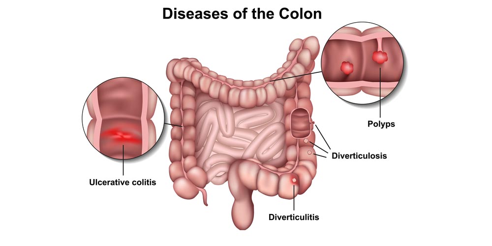
40 Colon diagram Vector Images, Colon diagram Illustrations - Depositphotos 40 Colon diagram Stock Vector Images, Royalty-free Colon diagram Drawings & Illustrations. VectorMine Crohns disease vector illustration. Labeled diagram with diagnosis. VectorMine Ulcerative colitis vector illustration. Labeled anatomical infographic.

Colon Picture Image on MedicineNet.com
Colon: Anatomy, histology, composition, function | Kenhub Structure of the large intestine (overview diagram) The colon makes up the longest part of the large intestine.It begins from the caecum at the ileocecal valve and ends in the rectum.The colon is about 1.5 meters long and frames the convolute of the small intestine in the abdominal cavity. However it can be shortened and lie quite flexibly in case of an incomplete rotation of the umbilical ...

Large Intestine Labeling Diagram | Quizlet
Cecum Histology Slide with Labeled Image and Diagram This taenia coli is not commonly found in all species. If you want to know more about the histological features of the colon, you may read the following article. Histological features of the colon with the labeled diagram and microscope image; You will learn the details histological features of the colon and compare it with the cecum microscope ...

Large intestine labeling Diagram | Quizlet
Colon Histology Slide with Labeled Diagram » AnatomyLearner >> The ... Jun 4, 2022 - The colon histology slide possesses four distinct layers like a typical tubular organ. Find taeniae coli and intestinal glands in the colon. Pinterest. Today. Explore. When autocomplete results are available use up and down arrows to review and enter to select. Touch device users, explore by touch or with swipe gestures.
Large Intestine. Vector & Photo (Free Trial) | Bigstock
Abdomen and digestive system anatomy: diagrams labeled - IMAIOS Colon: three diagrams of the face of the colon and rectum, with or without the mesentery of the colon and taenia coli as well as a detailed illustration of the right colonic flexure. Large intestine

Large intestine anatomy Diagram | Quizlet
Colon Anatomy - Medical Art Library The colon is divided into four parts: the ascending, transverse, descendingand sigmoid. The ascending and transverse colon meet at the right hepatic flexure(near the liver). The transverse and descending colon meet at the left splenic flexure(near the spleen). The large intestine has three bands of longitudinal muscle fibers called taeniae coli.

Large Intestine isolated on a white background. Medical ...
Illustration Picture of Anatomy - Colon - eMedicineHealth The colon is the largest part of the large intestine, extending from the cecum to the rectum. It is 5 feet long and its function is to reabsorb water from digested food and concentrate solid waste material, known as stool. The colon is made of several sections. The ascending colon travels up the right side of the abdomen.
Large Intestine Diagram Images – Browse 5,998 Stock Photos ...
How does the bowel work? Bowel information with diagrams ...

Colon anatomy: Pictures, features, and function

Colon Picture Image on MedicineNet.com

Large Intestine Anatomy, Parts, Diagram & Major Function ...

Large Intestine Anatomy, Parts, Diagram & Major Function ...

Large Intestine (Colon) - Diagram
SEER Training: Small & Large Intestine
Anatomy of the Colon and Rectum - Pristyn Care

Illustration Picture of Anatomy - Colon
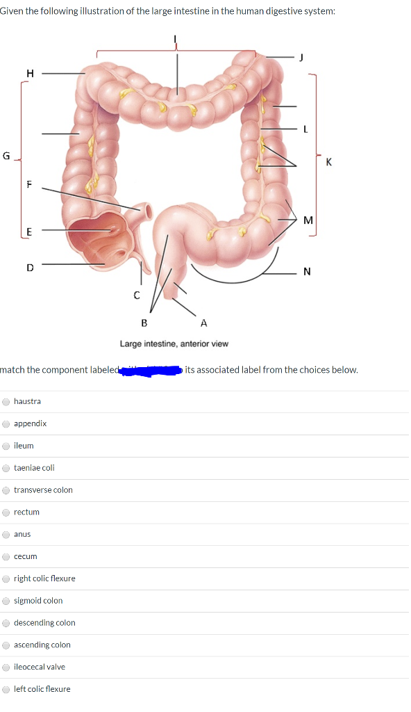
Solved Given the following illustration of the large | Chegg.com

Gastrointestinal tract 5: the anatomy and functions of the ...

Large Intestine - Location, Anatomy, Diagram, Structure ...

Large intestine structure | Large intestine diagram | Large ...
Colon Anatomy: Gross Anatomy, Microscopic Anatomy, Natural ...

Anatomy of the Large Intestine | Doctor Stock
The Colon - Ascending - Transverse - Descending - Sigmoid ...
Parts Colon Color Coded Labeled Stock Illustration 228843118 ...

Large Intestine Anatomy, Parts, Diagram & Major Function ...

unit 5 small and large intestine labeling Diagram | Quizlet

How Long Are Your Intestines? Length of Small and Large ...
![Anatomy and Physiology of the Large Intestine [Colon]](https://i.ytimg.com/vi/F4W2DLIKXww/maxresdefault.jpg)
Anatomy and Physiology of the Large Intestine [Colon]

Gastrointestinal tract 5: the anatomy and functions of the ...
Large Intestine Structure Mucosa and Musculature of Colon ...
Post a Comment for "38 colon diagram with labels"