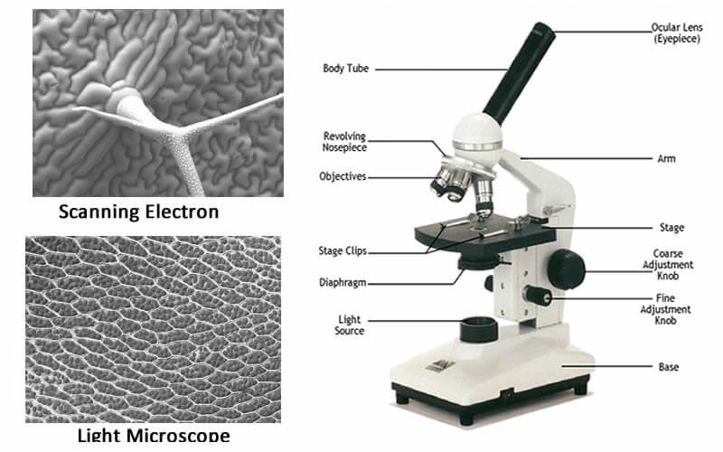
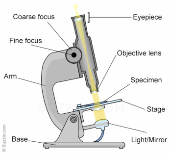
44 dissecting microscope diagram with labels
High-throughput identification and quantification of single … 22.02.2022 · Here, Jin et al., develop a method called Barcoding Bacteria for Identification and Quantification (BarBIQ), which allows to both characterize the global microbiome and to … Microscope labeled diagram - SlideShare 1. The Microscope Image courtesy of: Microscopehelp.com Basic rules to using the microscope 1. You should always carry a microscope with two hands, one on the arm and the other under the base. 2. You should always start on the lowest power objective lens and should always leave the microscope on the low power lens when you finish using it. 3.
rsscience.com › stereo-microscopeParts of Stereo Microscope (Dissecting microscope) - Rs' Science Stereo microscopes (also called Dissecting microscope) are branched out from other light microscopes for the application of viewing "3D" objects. These include substantial specimens, such as insects, feathers, leaves, rocks, sand grains, gems, coins, and stamps, etc. Functionally, a stereo microscope is like a powerful magnifying glass.

Dissecting microscope diagram with labels
The Rise and Future of Discrete Organic–Inorganic Hybrid … 28.05.2022 · Hybrid nanomaterials (HNs), the combination of organic semiconductor ligands attached to nanocrystal semiconductor quantum dots, have applications that span a range of practical fields, including biology, chemistry, medical imaging, and optoelectronics. Specifically, HNs operate as discrete, tunable systems that can perform prompt fluorescence, energy … › visual-impairmentsVisual Impairments | NSTA Use of a dissecting microscope and closed circuit television can help model during dissections. Be sure to familiarize the student with a visual impairment regarding the use of cutting instruments. (Videos are available online to instruct teachers about dissection with students who are visually impaired.) Group Interaction and Discussion Parts of the Dissecting Microscope - Synonym 6 Focus Knob The head of the microscope can be moved up and down with the focus knob, allowing the observer to view the image sharply; this is called rack and pinion focusing. 7 Stage Plate The specimen is placed on the stage plate for viewing. This plate is mounted on the base of the microscope, directly under the objective lens.
Dissecting microscope diagram with labels. Label the microscope — Science Learning Hub Use this with the Microscope parts activity to help students identify and label the main parts of a microscope and then describe their functions. Drag and drop the text labels onto the microscope diagram. If you want to redo an answer, click on the box and the answer will go back to the top so you can move it to another box. Parts of Dissecting Microscope | Botany - Biology Discussion In this article we will discuss about the parts of dissecting microscope with its working and utility. 1. Foot or Base: ADVERTISEMENTS: It is the basal, horse-shoe shaped or circular part of dissecting microscope. It is made of heavy material. It provides support to other parts of microscope. Compound Microscope Parts - Labeled Diagram and their Functions - Rs ... There are three major structural parts of a compound microscope. The head includes the upper part of the microscope, which houses the most critical optical components, and the eyepiece tube of the microscope. The base acts as the foundation of microscopes and houses the illuminator. The arm connects between the base and the head parts. › pmc › articlesAdvanced Fluorescence Microscopy Techniques—FRAP, FLIP, FLAP ... Apr 02, 2012 · 1. Introduction. FRAP, FLIP, FLAP, FRET, and FLIM are fluorescence microscopy techniques that in some way take advantage of particular aspects of the fluorescence process by which fluorochromes are excited and emit fluorescent light, are damaged during repetitive excitation, or undergo non-radiative decay prior to light emission.
Microscope Types (with labeled diagrams) and Functions These microscopes work on the principle called contrast-enhancing technique that is utilized to produce high-contrast images to view them with more accuracy and clarity. Phase-contrast microscope labeled diagram Phase-contrast microscope functions: Its applications areas include In cases where the specimen is colorless and is very tiny Everything You Need to Know About A Dissecting Microscope A dissecting microscope, or more commonly known as a stereo microscope, is a microscope that gives a three-dimensional view of a specimen. This is because of the binocular head, or the two eyepieces that are slightly angled, which creates the perfect peripheral vision that results in a three-dimensional visual. Compound Light/Dissecting Microscope Diagram | Quizlet Used to examine material mounted on microscope slides (usually thinly sectioned & stained) Provides total magnification of 40x-1000x No space for dissection Rules TRANSPORT Arm & base USE Always start at 4x, Coarse focus, Fine focus Then change objectives & use fine focus as needed Coarse focus ONLY with 4x! CLEANING Objectives/Oculars COVID-19 immune features revealed by a large-scale single-cell ... 01.04.2021 · Single-cell RNA sequencing (scRNA-seq) is powerful at dissecting the immune responses and has been applied to COVID-19 studies (Cao et al., 2020; Chua et al., 2020; Fan et al., 2020; Su et al., 2020; Wen et al., 2020; Xie et al., 2020; Zhang et al., 2020a, 2020b). While the current single-cell studies of COVID-19 have provided important cellular and molecular …
16 Parts of a Compound Microscope: Diagrams and Video Once you have an understanding of the parts of the microscope it will be much easier to navigate around and begin observing your specimen, which is the fun part! The 16 core parts of a compound microscope are: Head (Body) Arm. Base. Eyepiece. Eyepiece tube. Dissecting microscope (Stereoscopic or Stereo microscope) This microscope is a dual-powered dissecting microscope of 10x-30x with an ability to rotate 360° making it ideal for viewing and focussing better to view samples. By rotating the lenses, users can change the magnification of image. What is a Refractometer & How Does it Work - Cole-Parmer 05.03.2020 · What is a Refractometer? A refractometer is a simple instrument used for measuring concentrations of aqueous solutions. It requires only a few drops of liquid, and is used throughout the food, agricultural, chemical, and manufacturing industries.. How a Refractometer Works. When light enters a liquid it changes direction; this is called refraction. Dissecting Microscope Parts And Functions. All You Need To Know The dissecting microscope is also referred to as a stereoscopic microscope and is ordinarily used to study three-dimensional objects. And also as the name suggests for dissecting and analysing biological specimens under low magnification between two and two hundred and fifty times.
Binocular Microscope Anatomy - Parts and Functions with a Labeled Diagram Now, I will discuss the details anatomy of the light compound microscope with the labeled diagram. Why it is called binocular: because it has two ocular lenses or an eyepiece on the head that attaches to the objective lens, this ocular lens magnifies the image produced by the objective lens. Binocular microscope parts and functions
› tech-article › refractometersWhat is a Refractometer & How Does it Work - Cole-Parmer Mar 05, 2020 · Measurements are read at the point where the prism and solution meet. With a low concentration solution, the refractive index of the prism is much greater than that of the sample, creating a large refraction angle and a low reading ("A" on diagram). The reverse would happen with a high concentration solution ("B" on diagram).
Label the microscope Diagram | Quizlet Start studying Label the microscope. Learn vocabulary, terms, and more with flashcards, games, and other study tools.
SERS Tags for Biomedical Detection and Bioimaging 24.01.2022 · Furthermore, the laser spot of the Raman microscope is at the micrometer level (generally about 1 µm 2), in ... J. Interrogating cells, tissues, and live animals with new generations of surface-enhanced Raman scattering probes and labels. ACS Nano. 2017;11 :1136-1141 14. Ramya AN, Arya JS, Madhukrishnan M, Shamjith S, Vidyalekshmi MS, Maiti KK. …


Post a Comment for "44 dissecting microscope diagram with labels"